Acetyl-CoA fluorometric assay kit, Acetyl-CoA assay, Coenzyme A metabolism, fatty-acid synthesis, TCA cycle, mitochondrial metabolism, glycolysis-TCA linkage, acetyl-CoA quantification, fluorescent detection, metabolic flux.
Secondary keywords: CoA thioesters, acetyltransferase, citrate synthase, β-oxidation, PDH complex, ACSS2, metabolic reprogramming, NADH/NAD⁺ balance, sample preparation, LOD, linear range.
What is Acetyl-CoA and why measure it?
Acetyl-CoA is the central two-carbon donor in cell metabolism (linking glycolysis to the TCA cycle, fueling fatty-acid synthesis, cholesterol biosynthesis, protein acetylation, and parts of amino-acid catabolism). It sits at the crossroads of carbohydrate, lipid, and protein metabolism and is a key readout for metabolic flux in cancer biology, immunometabolism, stem-cell biology, and bioenergetics studies. Authoritative overviews and reference data can be found at the NIH/NCBI and NLM:
-
NCBI PubChem record for Acetyl-CoA (structure, identifiers, physicochemical data): https://pubchem.ncbi.nlm.nih.gov/compound/Acetyl-CoA
-
NLM MeSH entry for Acetyl-Coenzyme A: https://meshb.nlm.nih.gov/record/ui?ui=D000087
-
NCBI Bookshelf (metabolism & biochemistry textbooks index): https://www.ncbi.nlm.nih.gov/books/
-
NCBI Gene (e.g., ACSS2, a cytosolic/nuclear acetyl-CoA synthetase central to lipid synthesis and histone acetylation): https://www.ncbi.nlm.nih.gov/gene/55902
-
NIDDK (NIH) general resources on metabolism and metabolic pathways: https://www.niddk.nih.gov/
Why quantify acetyl-CoA? Because absolute/relative acetyl-CoA pools reflect pathway usage (e.g., glucose-derived acetyl-CoA via pyruvate dehydrogenase (PDH), acetate-derived via ACSS2, or fatty-acid-derived via β-oxidation). Tracking acetyl-CoA supports studies on growth signaling, epigenetic acetylation, mitochondrial health, and nutrient dependence.
Fluorometric assay principle (high-level)
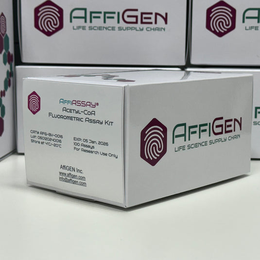
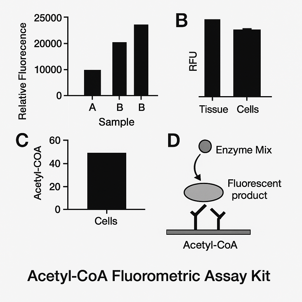
Most Acetyl-CoA Fluorometric Assay Kits use an enzyme cycling principle: free acetyl-CoA in the sample is converted enzymatically to a product that generates a fluorophore (often via an HRP- or oxidase-coupled reaction) proportional to acetyl-CoA concentration. The readout is typically Ex/Em ≈ 535/587–590 nm (kit-dependent).
-
Primer on enzyme-linked assays and readouts (NIH/NCBI): https://www.ncbi.nlm.nih.gov/pmc/
-
Fundamentals of fluorescence detection and linearity (NIST): https://www.nist.gov/programs-projects/fluorescence
-
Plate reader practices & instrument QA concepts (NIST): https://www.nist.gov/
Kits often include: Assay buffer, enzyme mix, developer/probe, Acetyl-CoA standard, and stop solution (varies by vendor). Assays are optimized to minimize interference from free CoA, CoA-thiols, and acetate; always consult the kit’s cross-reactivity table and recommended sample cleanup.
Sample types, storage, and pre-analytics
Typical matrices: mammalian cells, tissues (liver, muscle, brain), yeast, bacteria, and sometimes biofluids (if validated). Because acetyl-CoA is a labile thioester subject to hydrolysis and enzymatic turnover, cold, rapid extraction is essential.
Best practices (general, adopt to your kit SOP):
-
Quench metabolism rapidly (ice-cold buffers; work on ice).
-
Use protein-precipitation (e.g., PCA or perchlorate/methanol) or kit-specified extraction to remove enzymes that consume/produce acetyl-CoA.
-
Spin down debris; clarify supernatant.
-
Avoid repeated freeze–thaw. Aliquot at −80 °C.
Helpful institutional guidelines for lab handling & chemical hygiene:
-
NIH (environmental health & safety resources): https://ors.od.nih.gov/srm/Pages/default.aspx
-
CDC laboratory biosafety: https://www.cdc.gov/labs/
-
OSHA laboratory safety guidance: https://www.osha.gov/publications/laboratory-safety-guidance
-
EPA hazardous waste in labs: https://www.epa.gov/hwgenerators/management-hazardous-waste-laboratories
-
UC Berkeley EH&S chemical safety: https://ehs.berkeley.edu/
-
Cornell EHS lab safety: https://ehs.cornell.edu/
-
UMich EHS: https://ehs.umich.edu/
-
UC San Diego research safety: https://blink.ucsd.edu/safety/research-lab/
Assay workflow (typical 96-well plate format)
-
Standards & curve: Prepare serial dilutions of Acetyl-CoA standard (e.g., 0, 0.05, 0.1, 0.2, 0.5, 1, 2 µM; your kit may differ).
-
Samples: Add equal volumes to wells; keep total organic solvent content low to protect enzyme activity.
-
Reagent addition: Add enzyme mix and fluorogenic probe as directed.
-
Incubation: 20–60 min at room temperature or 37 °C (kit-specific). Protect from light.
-
Readout: Measure fluorescence (Ex/Em per kit).
-
Quantification: Fit a 4-parameter logistic (4PL) or linear model in the reliable range. Apply dilution factors.
General ELISA/plate-assay data-handling principles from NLM/NCBI and NIH:
-
Data analysis frameworks and QA in biomedical assays (NIH): https://www.nih.gov/
-
NCBI Bookshelf (statistics & methods compendia): https://www.ncbi.nlm.nih.gov/books/
Performance characteristics to document
-
Limit of detection (LOD) and Lower limit of quantitation (LLOQ) (often low-nanomolar to sub-µM depending on kit/instrument).
-
Dynamic range (typically ~10^2–10^3-fold).
-
Precision: Intra-assay CV and inter-assay CV (aim ≤10% where feasible).
-
Accuracy: Spike recovery (90–110%) and dilution linearity (±10%).
-
Specificity: Negligible response to CoA, acetyl-carnitine, citrate, ATP (verify in kit datasheet).
-
Matrix effects: Demonstrate equivalence across extraction buffers/tissues.
For validation and quality systems in regulated environments:
-
FDA quality system (device/IVD context; principles also help RUO labs): https://www.fda.gov/medical-devices/quality-system-qs-regulation
-
CLIA (clinical labs; if translating to CLIA-regulated work): https://wwwn.cdc.gov/clia/
-
NIST metrology principles (measurement assurance): https://www.nist.gov/topics/metrology
Interferences & controls
Potential interferences:
-
Endogenous thiols (GSH, CoA) and acetate (if the chemistry picks up acetyl donors) can bias signal if not controlled.
-
High NADH/NADPH in extracts can create autofluorescence (instrument-dependent).
-
Detergents, phenol red, and residual quenchers from extraction can attenuate signals.
Controls to include:
-
Blank (no acetyl-CoA).
-
No-enzyme control (probe only) to detect autofluorescence.
-
Spike-in recovery in each matrix.
-
Heat-inactivated sample to assess enzyme-dependent artifacts.
NIH/NLM technical resources on fluorometric assay caveats:
-
NCBI PMC (open-access methods & troubleshooting articles): https://www.ncbi.nlm.nih.gov/pmc/
-
NLM resources for assay development literature search: https://www.nlm.nih.gov/
Normalization strategies
Because acetyl-CoA content reflects cell number, protein mass, and mitochondrial density, normalize to:
-
Total protein (BCA/Bradford; dye compatibility with your assay needed).
-
Cell count (hemocytometer or automated counter).
-
Citrate synthase activity as a mitochondrial mass surrogate (many labs adopt this).
Background on mitochondrial content and metabolic readouts (NIH): https://www.nhlbi.nih.gov/
Biological interpretation
High acetyl-CoA may indicate:
-
Strong glucose oxidation (PDH flux).
-
Robust acetate utilization (ACSS2-driven, often under hypoxia or lipid anabolism).
-
Increased β-oxidation (fat-derived acetyl-CoA).
Low acetyl-CoA may reflect PDH inhibition (e.g., PDH phosphorylation), ETC impairment, limited substrate supply, or rapid consumption by citrate synthase or acetyltransferases (e.g., histone acetylation).
Useful NIH program pages for metabolism & disease:
-
NCI cancer metabolism: https://www.cancer.gov/research/areas/biology/metabolism
-
NIDDK metabolic disease resources: https://www.niddk.nih.gov/
-
NIAID immunometabolism basics: https://www.niaid.nih.gov/
Applications
-
Cancer metabolism: track re-routing to lipid synthesis, acetate scavenging (ACSS2-dependent). NCI background: https://www.cancer.gov/
-
Neuroscience/brain energy: evaluate shifts in acetyl-CoA pools during neuronal activity or glial metabolism (NIH BRAIN): https://braininitiative.nih.gov/
-
Immunometabolism: T-cell/macrophage activation alters acetyl-CoA and histone acetylation (NIAID): https://www.niaid.nih.gov/
-
Metabolic engineering & microbiology: quantify acetyl-CoA to optimize biosynthetic pathways (DOE/OSTI): https://www.osti.gov/
-
Mitochondrial disease models: link acetyl-CoA to PDH/ETC function (NIH: National Institute of Neurological Disorders and Stroke): https://www.ninds.nih.gov/

Step-by-step protocol (template; adapt to your kit)
-
Prepare standards in assay buffer (follow kit range).
-
Extract samples rapidly on ice using kit-recommended buffer or a validated organic extraction; neutralize if required.
-
Plate layout: load standards, blanks, samples (≥ duplicates).
-
Add enzyme mix and probe (light-sensitive).
-
Incubate 30–45 min at RT or 37 °C; avoid bubbles and plate edge effects.
-
Read fluorescence (Ex/Em per kit).
-
Calculate concentrations via standard curve (4PL recommended if curve sigmoidal).
-
Apply dilution factor and normalize (e.g., to mg protein).
-
Record CVs, spike recovery, linearity.
-
Archive raw plate files for QA (FDA/GLP mindset): https://www.fda.gov/science-research/good-laboratory-practices-glp
Troubleshooting guide
| Issue | Likely cause | Corrective action |
|---|---|---|
| Weak signal | Low analyte, probe decay, improper Ex/Em | Use fresh probe; confirm instrument filters; concentrate sample |
| High background | Autofluorescence, inadequate washing/cleanup | Add no-enzyme controls; optimize extraction; reduce matrix fluorescence |
| Non-linear standards | Pipetting error, wrong fit model | Re-prepare standards; use 4PL; ensure linear region for interpolation |
| High CVs | Edge effects, bubbles, inconsistent timing | Avoid edge wells; spin plates briefly; standardize incubation |
| Matrix interference | Detergents/thiols quenching | Further cleanup; verify spike recovery; dilute sample |
General biosafety & chemical hygiene references:
-
OSHA lab guidance: https://www.osha.gov/publications/laboratory-safety-guidance
-
NIH biosafety policies: https://osp.od.nih.gov/biotechnology/biosafety/
Reporting & data integrity (publication/QA)
-
Report LOD/LLOQ, dynamic range, CVs, recovery, linearity, n, normalization.
-
Provide plate map, standard curve, and instrument settings.
-
Document sample extraction and storage.
-
Align with CLIA/good-measurement principles where applicable: https://wwwn.cdc.gov/clia/ and NIST metrology: https://www.nist.gov/topics/metrology
Safety and waste
Follow your institution’s chemical hygiene plan and local regulations for disposal of organic extracts and oxidizing substrates. Useful anchors:
-
EPA lab hazardous waste: https://www.epa.gov/hwgenerators/management-hazardous-waste-laboratories
-
NIH occupational safety programs: https://ors.od.nih.gov/srm/Pages/default.aspx
-
UC Berkeley EH&S: https://ehs.berkeley.edu/
Summary (for fast SEO snippets)
The Acetyl-CoA Fluorometric Assay Kit provides sensitive, quantitative measurement of acetyl-CoA in cells and tissues using an enzyme-coupled fluorescent reaction. With rapid extraction, robust standard-curve fitting, and careful control of interferences, researchers can monitor TCA-cycle input, lipogenesis, acetate utilization, and epigenetic acetylation potential. Validation should include LOD/LLOQ, CVs, recovery, linearity, and matrix checks to ensure dependable data—principles consistent with FDA, CDC/CLIA, NIH, and NIST best practices.
Useful .gov / .edu references (embedded above; collected here)
-
NCBI PubChem Acetyl-CoA: https://pubchem.ncbi.nlm.nih.gov/compound/Acetyl-CoA
-
NLM MeSH Acetyl-Coenzyme A: https://meshb.nlm.nih.gov/record/ui?ui=D000087
-
NCBI Bookshelf (biochemistry texts): https://www.ncbi.nlm.nih.gov/books/
-
NCBI Gene ACSS2: https://www.ncbi.nlm.nih.gov/gene/55902
-
NIDDK (metabolism resources): https://www.niddk.nih.gov/
-
NIST (fluorescence & measurement science): https://www.nist.gov/programs-projects/fluorescence
-
NIH Office of Research Services (safety): https://ors.od.nih.gov/srm/Pages/default.aspx
-
CDC Laboratory Programs: https://www.cdc.gov/labs/
-
OSHA Laboratory Safety: https://www.osha.gov/publications/laboratory-safety-guidance
-
EPA Hazardous Waste (labs): https://www.epa.gov/hwgenerators/management-hazardous-waste-laboratories
-
UC Berkeley EH&S: https://ehs.berkeley.edu/
-
Cornell EHS: https://ehs.cornell.edu/
-
UMich EHS: https://ehs.umich.edu/
-
UC San Diego Research Safety: https://blink.ucsd.edu/safety/research-lab/
-
NCI Cancer Metabolism: https://www.cancer.gov/research/areas/biology/metabolis