Introduction
Fibronectin ELISA (Enzyme-Linked Immunosorbent Assay) is a robust analytical tool used for the quantitative measurement of fibronectin — a high-molecular-weight extracellular matrix glycoprotein essential for cell adhesion, migration, wound healing, and tissue repair.
Fibronectin exists in soluble plasma form and insoluble cellular form, both playing key roles in embryogenesis, angiogenesis, and oncogenic progression. According to the National Center for Biotechnology Information (NCBI), fibronectin acts as a structural scaffold connecting collagen, fibrin, and integrins, and modulating the cellular cytoskeleton.
The Fibronectin ELISA assay quantitatively measures fibronectin concentrations in serum, plasma, tissue homogenates, or cell culture supernatants through specific antibody–antigen interactions, providing valuable insight for biomedical diagnostics, fibrosis studies, and cancer biomarker profiling.
![AffiELISA® Human Fibronectin ELISA [ FN1]](https://affigen.com/cdn/shop/files/5BAFG-E4345_5D_20AffiELISA_C2_AE_20Cattle_20IFNg_20Kit_20High-Resolution_20Interferon_20Gamma_20ELISA_20Detection_6407516e-2cb2-47e2-a488-85ac447ff0eb_535x.png?v=1712843239)
Structural and Biological Overview of Fibronectin
Fibronectin is a dimeric glycoprotein (~440 kDa) composed of two nearly identical subunits linked by disulfide bonds at the C-terminus.
Each subunit includes type I, II, and III repeats, which enable binding to collagen, fibrin, heparin, and cell surface integrins (e.g., α5β1, αvβ3) (Protein Data Bank – PDB).
The RGD sequence (Arg-Gly-Asp) in the type III domain is crucial for cell adhesion and signal transduction, as established by structural biology studies at the National Institutes of Health (NIH).
Fibronectin plays critical physiological roles in:
-
Wound healing — scaffolding for fibroblast migration.
-
Cell differentiation and morphogenesis — influencing ECM remodeling (PubMed – NIH).
-
Oncology — elevated fibronectin correlates with tumor metastasis and angiogenesis.
-
Fibrosis — accumulation in liver, lung, and renal fibrotic tissues.
For molecular details, consult NCBI Gene Database for FN1 (Fibronectin 1) gene encoding human fibronectin.
Principle of Fibronectin ELISA
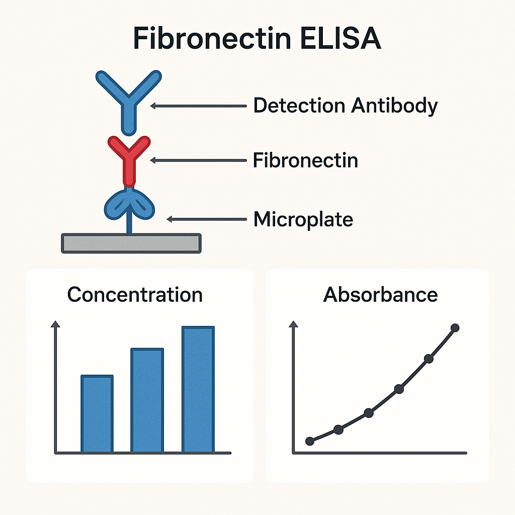
The Fibronectin ELISA employs the antigen–antibody reaction to detect fibronectin concentration with high specificity. The assay can be performed in sandwich or indirect formats, depending on the antibody pair used.
a. Sandwich ELISA:
-
Capture antibody is pre-coated on a 96-well microplate.
-
Sample or standard containing fibronectin binds to the capture antibody.
-
Biotinylated detection antibody binds the captured fibronectin.
-
Streptavidin–HRP conjugate amplifies the signal.
-
Addition of TMB substrate yields a colorimetric reaction proportional to fibronectin concentration.
b. Detection Wavelength: 450 nm, as recommended by the FDA Analytical Procedure Guidelines.
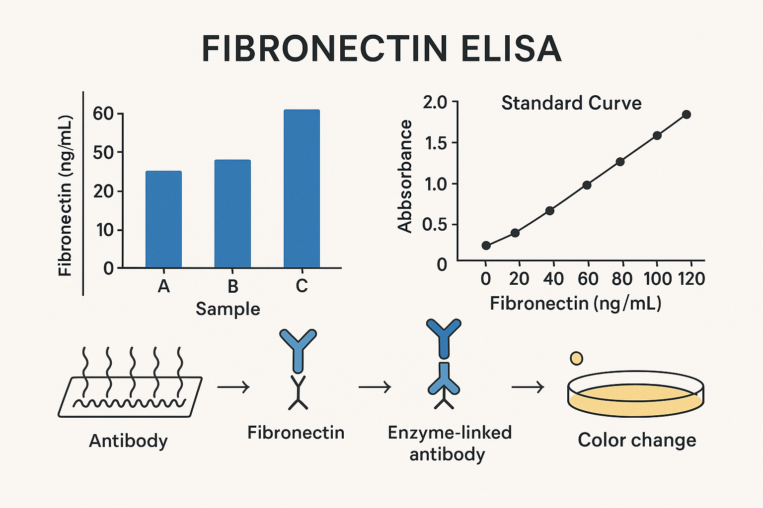
The color intensity (optical density) is measured with a microplate reader, and fibronectin concentration is determined via a standard calibration curve (CDC Laboratory Reference).
Sample Preparation and Handling
Proper sample processing ensures assay reproducibility and accuracy. The National Institute of Standards and Technology (NIST) advises adherence to Good Laboratory Practices (GLP) for sample integrity.
| Sample Type | Preparation Steps |
|---|---|
| Plasma/Serum | Collect in EDTA or heparin tubes, centrifuge 15 min at 3000 rpm, store at –80°C. |
| Cell culture supernatant | Remove debris by centrifugation; filter through 0.22 µm membrane. |
| Tissue lysate | Homogenize in PBS + protease inhibitors; clarify by centrifugation. |
Avoid repeated freeze–thaw cycles, which can degrade glycoprotein epitopes (NIH Laboratory Safety Manual).
Standard Curve and Quantification
Fibronectin concentration is calculated by interpolating the sample OD values on a standard curve generated with known fibronectin standards (0–2000 ng/mL).
According to NIST ELISA Calibration Recommendations, linear regression or 4-parameter logistic (4PL) models are used for curve fitting.
Each plate should include blank, negative control, and quality control samples to verify assay precision and linearity.
Reagents and Materials
-
96-well microplate pre-coated with anti-human fibronectin antibody
-
Standards: recombinant human fibronectin
-
Detection antibody: biotinylated anti-fibronectin IgG
-
Streptavidin–HRP conjugate
-
TMB substrate and stop solution (H₂SO₄ 2N)
-
Wash buffer (PBS-T) and assay diluent
Manufacturing and quality control standards are based on U.S. Pharmacopeia (USP) and FDA Biologics Regulations.
Mechanism of Signal Detection
The colorimetric signal arises from HRP (horseradish peroxidase)-mediated oxidation of TMB substrate in the presence of hydrogen peroxide.
The enzymatic reaction kinetics are described in detail by the National Library of Medicine (PubChem).
Absorbance is read at 450 nm within 15 minutes of stopping the reaction, ensuring accuracy and avoiding chromophore degradation (NIH Biomedical Instrumentation Center).
Biological and Clinical Relevance of Fibronectin Quantification
Elevated fibronectin levels serve as biomarkers in multiple physiological and pathological states:
| Condition | Fibronectin Expression | Clinical Insight |
|---|---|---|
| Wound healing | Upregulated | Indicates matrix remodeling (PubMed) |
| Sepsis | Reduced plasma fibronectin | Predictive of poor prognosis (CDC Clinical Guidelines) |
| Cancer metastasis | Increased expression | Associated with tumor invasion (NIH Cancer Research) |
| Fibrosis (liver/lung) | Elevated | Marker of ECM accumulation (NIH/NIDDK Liver Research) |
| Diabetic nephropathy | Elevated urinary fibronectin | Indicator of glomerular injury (NIDDK Kidney Disease Info) |
Applications of Fibronectin ELISA
a. Biomedical Diagnostics
Used to quantify fibronectin in human serum and plasma for research in fibrotic diseases, vascular disorders, and oncology.
The FDA CBER lists fibronectin as a biological quality marker in blood plasma studies.
b. Tissue Engineering and Regenerative Medicine
Fibronectin levels are assessed to monitor cell–matrix interactions and biomaterial compatibility, according to NIH Tissue Engineering Branch.
c. Cell Culture Studies
Quantifying fibronectin secreted by fibroblasts or epithelial cells provides insights into extracellular matrix formation, as reported by Johns Hopkins Cell Biology Department.
d. Pharmaceutical and Biomarker Validation
Fibronectin ELISA assays are used in drug discovery pipelines for assessing the anti-fibrotic efficacy of compounds (NCBI – Drug Screening Database).
Troubleshooting and Optimization
| Problem | Possible Cause | Solution |
|---|---|---|
| Low signal | Insufficient antibody binding | Extend incubation or increase antibody concentration |
| High background | Inadequate washing | Increase wash cycles (≥5x) |
| Nonlinear standard curve | Improper dilution | Prepare fresh serial dilutions |
| High variability | Pipetting error | Use calibrated pipettes, replicate wells |
| Edge effect | Uneven plate temperature | Use consistent incubation conditions |
Best practices for assay optimization are outlined in NIH Reproducibility Guidelines.
Safety and Handling
All human samples must be treated as potentially infectious following BSL-2 containment as per CDC Biosafety in Microbiological Laboratories.
Chemical reagents should be handled using PPE and under fume hoods, following Occupational Safety and Health Administration (OSHA) standards.
Data Analysis and Interpretation
Optical density (OD) data are plotted using log-log or 4PL regression models.
Inter-assay and intra-assay coefficients of variation (CV%) should be <10% to ensure assay precision (NIST Bioprocess Quality Metrics).
The results can be normalized to total protein concentration or cell number depending on experimental design (NIH Proteomics Standards).
Advantages of Fibronectin ELISA
| Advantages | Details |
|---|---|
| High sensitivity and specificity | Dual-antibody capture and detection |
| Quantitative results | Direct correlation between OD and fibronectin concentration |
| Wide dynamic range | 0.1–2000 ng/mL |
| Compatibility | Serum, plasma, tissue, and cell culture |
| Reproducibility | Standardized under GLP and ISO 13485 |
Comparative ELISA assay validations are documented by the FDA Biologics Evaluation Report.
Limitations
-
Requires strict temperature control (18–25 °C)
-
Not suitable for fibronectin fragments <30 kDa
-
Cross-reactivity with homologous ECM proteins (e.g., vitronectin) may occur
-
Limited stability of reconstituted standards (≤2 weeks at 4°C)
Refer to the NIH Bioassay Quality Guidelines for troubleshooting.
Emerging Research and Future Directions
Recent advances have enhanced Fibronectin ELISA assays with:
-
Fluorometric and chemiluminescent detection systems for femtogram sensitivity (NIST Light Measurement Lab).
-
Microfluidic ELISA-on-chip for rapid, low-volume testing (NIH Bioengineering and Nanomedicine).
-
Multiplexed immunoassays detecting fibronectin alongside laminin and collagen IV in ECM panels (NCBI Proteomics Studies).
-
AI-based data interpretation to reduce intra-lab variability and improve quantification accuracy.
Conclusion
The Fibronectin ELISA remains a cornerstone technique for evaluating extracellular matrix dynamics, fibrosis progression, and tissue remodeling in both research and clinical settings.
Its high sensitivity, quantitative accuracy, and reproducibility make it a preferred choice for studies of wound healing, tumor biology, and organ fibrosis.
By combining antibody-based specificity with robust signal amplification, Fibronectin ELISA continues to advance the precision of molecular diagnostics and translational research in cell biology and biomedical science.